Tissue Collection
For the purposes of our investigation, ovaries were collected from the Greeley slaughter house and transferred to University of Northern Colorado Greeley in 1x PBS. When the ovaries arrived at the university the Corpus Luteum (CL) was dissected away from the ovary. The CL was cut into 1 mm slices and placed in a solution called Hank’s Balanced Salt Solution(HBSS). Then the samples were moved to T75 culture flasks and incubated in a rocking water bath at 37°C with type 1 collagenase and Bovine Serum Albumin (BSA). The samples then incubated for a few hours in a rocking water bath at 37°C. After incubation the samples were pushed through a filter to separate the larger debris, such as connective tissue. Then the samples were spun down in a centrifuge at 500 G, and transferred to T75 culture flasks and resuspended in HAMS-F12 media, FBS, insulin transferrin serum, and antiviral-antibacterial supplements (Anti Anti).(8)
Flow Cytometry
Cells were then prepared for flow cytometry by removing the culture media from the T75 culture flasks. Each flask was washed two times with 1x PBS. After that, approximately 4 mL of trypsin is added to each flask, then the cells incubated with the trypsin at 37°C for 15-20 minutes. The trypsin and cell mixture was then pipetted into a 15 mL conocal tube and spun down in a centrifuge at 500 G for 5 minutes. The supernatant was removed and the pellet was suspended in 1% BSA-PBS. The cells were divided into microcentrifuge tubes according to their corresponding treatment groups. The first treatment group received 1% PBS and BSA, while the second treatment group received TMRM (tetramethylrhodamine, concentrated at 100 nanomolar) added to 1% BSA. Finally, the third treatment group was incubated with CCCP (Carbonyl cyanide m-chlorophenyl hydrazone) with a 50 mM stock concentration) to form the negative control group. The cells treated with CCCP then incubated at 37°C for five minutes, after which TMRM was added to them. (8) After this, they are ran by the flow cytometer for graphs.
Confocal Microscopy
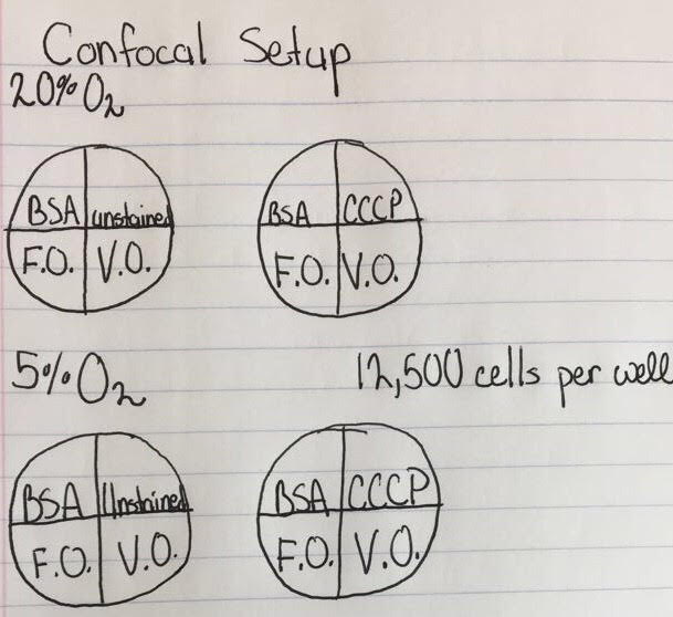
Specific groups were ran for our Confocal Microscopy, including a 20% oxygenated environment where there were two four-well plates and a 5% oxygenated environment where there were also two four-well plates. Each plate in each hypoxic environment contained a vegetable oil treatment group, a BSA control treatment group, a CCCP treatment group, and a fish oil treatment group. A cell sorter was utilized to deliver exactly 12,500 cells into each well. (8)
The vegetable oil and fish oil were bound to BSA by incubation in a rocking water bath for two hours at 37°C before being applied as a treatment. After being bound they were then applied to their specific groups. CCCP had not yet been applied. These cells were then incubated for three days at 37°C and then grown in either 20% oxygen or 5% oxygen for 24 hours. At hour 24 of this study 0.5 microliters of TMRM was applied to the BSA control, fish oil, and vegetable oil groups. Then the CCCP was applied to it’s designated cells. After this the cells were observed under the confocal microscope. (8)
